
Efficacy of Jaloukavacharana (Leech Therapy) and Kasisadi Taila in the management of Venous Leg Ulcer - A Single Case Report
DOI:
https://doi.org/10.21760/jaims.10.7.55Keywords:
Ayurveda, Jaloukavacharana, Kasisadi Taila, Pippali Churna, Haritaki Churna, Venous leg ulcerAbstract
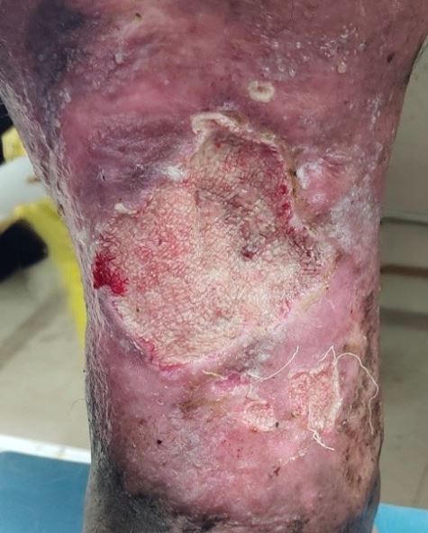
Venous leg ulcers are common chronic wounds resulting from impaired venous return in the lower limbs. They often reduce quality of life due to pain, discharge, restricted mobility, and the risk of infection. Although conventional treatments such as medical therapy, wound dressing, and surgery are available, the recurrence rate remains high. Ayurveda offers promising alternatives, particularly Jaloukavacharana (leech therapy), which facilitates wound healing through the pharmacologically active constituents in leech saliva, including anticoagulants, vasodilators, and antimicrobial agents. Kasisadi Taila, an Ayurvedic medicated oil, is also traditionally used for its wound-cleansing and healing properties, although clinical documentation is limited. This case report presents a 48-year-old non-diabetic male with a chronic, non-healing ulcer on the right lower leg associated with varicose veins. The patient underwent Jaloukavacharana once weekly, followed by dressing with Kasisadi Taila. Internal medications included Pippali Churna (three gram twice daily before food with warm water) to enhance digestion and circulation, and Haritaki Churna (five gram at bedtime with warm water) to support bowel regularity. The ulcer showed progressive improvement, with complete healing observed after eleven weeks. This case suggests that the integrated use of Jaloukavacharana, Kasisadi Taila, and supportive Ayurvedic oral medications can be an effective and safe alternative in managing venous leg ulcers. The combination not only promoted wound healing but also prevented complications without any adverse effects. Further clinical studies are recommended to validate these findings on a larger scale.
Downloads
References
O'Donnell TF Jr, Passman MA, Marston WA, et al. Management of venous leg ulcers: clinical practice guidelines of the Society for Vascular Surgery® and the American Venous Forum. J Vasc Surg. 2014;60(2 Suppl):3S–59S. Available from: https://www.jvascsurg.org/article/S0741-5214(14)00851-9/fulltext. doi:10.1016/j.jvs.2014.04.049
Cushman M. Epidemiology and risk factors for venous thrombosis. Semin Hematol. 2007;44(2):62–69. Available from: https://pubmed.ncbi.nlm.nih.gov/17433897/. doi:10.1053/j.seminhematol.2007.02.004
Abbade LPF, Lastória S. Venous ulcer: epidemiology, physiopathology, diagnosis and treatment. Int J Dermatol. 2005;44(6):449–456. Available from: https://onlinelibrary.wiley.com/doi/10.1111/j.1365-4632.2004.02456.x. doi:10.1111/j.1365-4632.2004.02456.x
Prakash S, Tiwary SK, Mishra M, Khanna AK. Venous ulcer: review article. Surg Sci. 2013;4(2):63–72. Available from: https://www.researchgate.net/publication/236163480_Venous_Ulcer_Review_Article. doi:10.4236/ss.2013.42028
Jindal R, Dekiwadia DB, Krishna PR, et al. Evidence-based clinical practice points for the management of venous ulcers. Indian J Surg. 2018;80(2):183. Available from: https://pubmed.ncbi.nlm.nih.gov/29915484/. doi:10.1007/s12262-018-1726-3
Thakral KK. Arsha Chikitsa. In: Sushrutha Samhitha, Chikitsa Sthana. Varanasi: Chaukhambha Sanskrit Sansthan; 2020. p. 268.
Ligi D, Mosti G, Croce L, Raffetto JD, Mannello F. Chronic venous disease – Part II: Proteolytic biomarkers in wound healing. Biochim Biophys Acta. 2016;1862(10):1900–1908. Available from: https://pubmed.ncbi.nlm.nih.gov/27460704/. doi:10.1016/j.bbadis.2016.07.011
Abbade LP, Lastória S. Venous ulcer: epidemiology, physiopathology, diagnosis and treatment. Int J Dermatol. 2005;44(6):449–456. Available from: https://pubmed.ncbi.nlm.nih.gov/15941430/. doi:10.1111/j.1365-4632.2004.02456.x
Amarprakash P, Dwivedi. Case study of leech application in diabetic foot ulcer. Int J Res Ayurveda Pharm. 2012;3(5):748–751. Available from: https://www.researchgate.net/publication/269846710_Case_study_of_leech_application_in_diabetic_foot_ulcer. doi:10.7897/2277-4343.03536
Sig AK, Guney M, Uskudar Guclu A, Ozmen E. Medicinal leech therapy—an overall perspective. Integr Med Res. 2017;6(4):337–343. Available from: https://pubmed.ncbi.nlm.nih.gov/29296560/. doi:10.1016/j.imr.2017.08.001
Kiran K, Asad M. Wound healing activity of Sesamum indicum L seed and oil in rats. Indian J Exp Biol. 2008;46(11):777–782. Available from: https://pubmed.ncbi.nlm.nih.gov/19090349/
Bhavamishra. Bhavaprakash, Purvakhanda 6/4/82–84. In: Prof KR, translator. Reprint ed. Varanasi: Chaukhamba Krishnadas Academy; 2016. p. 240.
Wlaschek M, Scharffetter-Kochanek K. Oxidative stress in chronic venous leg ulcers. Wound Repair Regen. 2005;13(5):452–461. Available from: https://pubmed.ncbi.nlm.nih.gov/16176453/. doi:10.1111/j.1067-1927.2005.00065.x
Mittal R, Gupta RL. In vitro antioxidant activity of piperine. Methods Find Exp Clin Pharmacol. 2000;22(5):271–274. Available from: https://pubmed.ncbi.nlm.nih.gov/11031726/. doi:10.1358/mf.2000.22.5.79664
Afshari AR, Sadeghnia HR, Mollazadeh H. A review on potential mechanisms of Terminalia chebula in Alzheimer's disease. Adv Pharmacol Sci. 2016;2016:8964849. Available from: https://pubmed.ncbi.nlm.nih.gov/26941792/. doi:10.1155/2016/8964849
Published
How to Cite
Issue
Section
License


